
The nervous system is classified into the central and peripheral nervous systems. Regarding the central nervous system, the functions of the brain are briefly explained, covering the forebrain (cerebrum and diencephalon), midbrain, and hindbrain (pons, medulla oblongata, and cerebellum), without delving into the brain’s internal structures and functions. The spinal cord’s functions, the reflex arc, and the importance of reflexes in human life are also emphasized.
In the peripheral nervous system, the general characteristics of the somatic and autonomic nervous systems are provided, but distinctions between sympathetic and parasympathetic nerves are not discussed.
While explaining the structure of the central and peripheral nervous systems, various visual elements (such as photographs, illustrations, drawings, and cartoons), graphic organizers (including concept maps, mind maps, and diagrams), and e-learning tools and applications (such as animations, videos, simulations, infographics, augmented and virtual reality applications) are utilized.
A reading passage is provided about Ibn Sina’s contributions to human physiology.
The nervous system is divided into two main sections: the central nervous system and the peripheral nervous system.
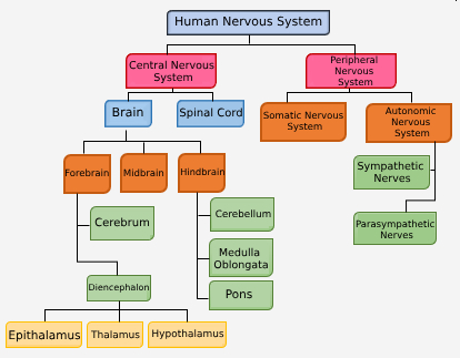
The central nervous system in humans is divided into two main parts: the brain and the spinal cord.
The brain and spinal cord receive and process various stimuli from both the internal and external environments.
The structure of the central nervous system consists of the cell bodies of motor neurons and interneurons.
A. Brain
- It is the command center of the body.
- In humans, the brain is located within the skull and protected by cranial bones.
- The brain weighs approximately 1,300–1,400 g.
- It consists of approximately 100 billion neurons and a significantly higher number of supporting cells.
The volume and mass of the brain are not related to intelligence and learning capacity. A larger surface area is one of the factors that make the human brain more developed than that of other vertebrates.
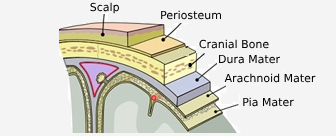
The brain is surrounded by a three-layered membrane called the meninges.
The meninges layers, from outermost to innermost, are called the dura mater, arachnoid mater, and pia mater.
- Dura mater: Located just beneath the skull, it protects the brain from mechanical impacts, injuries, and trauma.
- Arachnoid mater: Positioned between the dura mater and pia mater, it connects these layers with delicate connective tissue fibers resembling a spider web.
- Pia mater: The innermost layer of the meninges, it contains blood vessels that supply the brain with nutrients and oxygen.
The fluid that leaks from capillaries due to the effect of blood pressure forms the cerebrospinal fluid (CSF) and fills the space between the pia mater and the arachnoid mater.
The functions of cerebrospinal fluid (CSF):
- It protects the brain and spinal cord from mechanical impacts, such as blows and shocks.
- It facilitates the exchange of substances between blood and nerve cells.
- It helps maintain the balance of ion concentrations in the central nervous system.
If the cerebrospinal fluid becomes infected, it can lead to inflammation of the meninges, causing a disease known as meningitis.
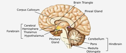
The brain consists of three main sections: the forebrain, midbrain, and hindbrain.
a. Forebrain
- Forebrain (Prosencephalon): This is the largest part of the brain. It is divided into two sections: the telencephalon (cerebral cortex) and diencephalon.
a. Telencephalon (Cerebrum): It consists of two parts, the left and right hemispheres. It covers the other parts of the brain from the top.
- When a cross-section is taken, the outer layer contains gray matter (cell bodies (soma)) in gray color, and the inner part contains white matter (the axons) in white color. The gray matter is known as the cerebral cortex.
- The brain hemispheres are connected by bundles of axons. These bundles are:
- The corpus callosum, which connects the hemispheres from the top.
- The brainstem connects the brain to the spinal cord.
- The groove separating the two hemispheres transversely is called the Longitudinal Fissure.
- Each hemisphere of the brain controls the opposite side of the body. Since approximately 90% of people have the left hemisphere dominant, the majority of people use their right hand. If damage occurs to the left hemisphere, the right hemisphere may develop dominance.
The cerebral hemispheres are the regions where centers for voluntary movement control, perception of stimuli from the five senses, learning, memory, intelligence, consciousness, writing, and speech are located.
A pigeon with its cerebral cortex removed
- Can walk if pushed
- Cannot fly unless stimulated
- Can fly if thrown into the air
- Does not feel hunger and does not eat the food placed in front of it
- Swallows food when placed in the pharynx
- Shows no response when a cat or dog approaches
- Is unresponsive to external stimuli
It has been observed that all of this animal’s movements are unconscious.
The size of the centers located in the cerebral cortex is directly proportional to the number and density of the receptors that send stimuli to this region.
The cerebral hemispheres are also divided into four regions that control different activities. These are:
- Frontal lobe
- Parietal lobe
- Temporal lobe
- Occipital lobe
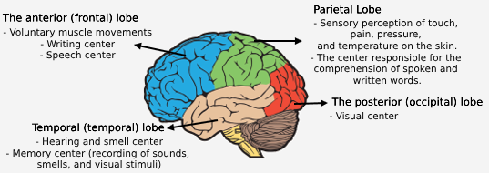
In case of damage to the temporal lobe, the person can notice the object but cannot recognize it.
b. Diencephalon: It is one of the parts that form the forebrain. It includes the epithalamus, thalamus, and hypothalamus regions.
- Epithalamus: It is located in the upper posterior part of the thalamus. It works together with the thalamus and hypothalamus. A fine extension of the epithalamus is called the pineal gland. The melatonin hormone secreted by this gland is known to affect biological rhythms related to reproduction. Additionally, some capillary networks that form the cerebrospinal fluid are located here.
- Thalamus: It is the central station for all senses, except for the sense of smell, where sensory signals are collected and distributed. Sensory signals are classified here and then transmitted to the sensory centers in the cerebral cortex. The thalamus regulates sleep and wakefulness states through impulses from other regions of the brain. (It facilitates the transition from wakefulness to sleep.)
- Hypothalamus: Its main functions include:
- Controlling the autonomic nervous system.
- Serving as the central regulation center for homeostasis.
- Regulating the function of the pituitary gland, which controls hormones secreted by other organs.
- Regulating body temperature, blood pressure, hunger, and sexual impulses.
Shivering is stimulated by the hypothalamus. The most intense shivering can increase body heat production by 4-5 times.
In cold-blooded animals, the hypothalamus is not well developed.
- It helps us experience emotions such as anger and pleasure.
- It functions as an “internal timer” for daily biological rhythms, such as sleep cycles and hunger.
- It regulates thirst, urine formation, and electrolyte balance.
- It regulates carbohydrate and fat metabolism.
- It controls the body’s water balance through the ADH hormone.
c. Midbrain: Located above the pons, it lies between the cerebellum and the diencephalon. It serves as a bridge between the forebrain and hindbrain.
- The midbrain controls visual and auditory reflexes. For example, the constriction of pupils in light and the erection of a dog’s ears in response to sound are regulated by these centers.
- Additionally, centers that regulate muscle tone (the slight contraction of muscles even at rest) and posture are also located in the midbrain.
d. Hindbrain: It consists of the cerebellum, medulla oblongata, and pons.
- Cerebellum: Located at the lower back of the brain, above the medulla oblongata. Like the brain, it is divided into two hemispheres. The cerebellar hemispheres are connected to each other by the pons. The outer part consists of gray matter, while the inner part contains white matter. The white matter branches into the gray matter, resembling a tree, and is therefore referred to as the “Arbor Vitae“.

The function of the cerebellum is to regulate muscle movements and maintain body balance. The cerebellum’s function is influenced by stimuli coming from the inner ear and eyes.
It has been experimentally observed that a dog with its cerebellum removed cannot walk. Babies cannot sit, stand, or walk before the cerebellum has fully developed. A bird with a damaged cerebellum cannot fly even if it flaps its wings when thrown into the air, as it cannot coordinate the movement of its wings.
A human whose cerebellum is damaged or surgically removed:
- Struggles to bring the fingers of both hands together.
- Cannot walk on a tightrope.
- Cannot write any word after picking up a pencil.
Medulla Oblongata (Hindbrain): Unlike the forebrain and cerebellum, it is composed of white matter on the outside and gray matter on the inside. It is a continuation of the spinal cord.
The motor nerves that exit from the cerebral hemispheres and travel to the body cross at the medulla oblongata. As a result, the nerves coming from the right hemisphere control the left side of the body, while the nerves coming from the left hemisphere control the right side of the body.
The medulla oblongata regulates the functioning of systems such as digestion, respiration, circulation, and excretion, and controls vital reflexes such as swallowing, sneezing, coughing, and vomiting.
Damage to the medulla oblongata results in the cessation of vital functions. For this reason, the medulla oblongata is also referred to as the “Center for vital activities.”
Pons (Pons Varolii): A part of the hindbrain, the pons consists of thick nerve bundles located between the midbrain and the medulla oblongata.
It connects the cerebellar hemispheres and facilitates the transmission of impulses between them.
Together with the medulla oblongata, it plays a role in respiration.
The pons, medulla oblongata, and midbrain together are referred to as the brainstem.
- The pons is found only in mammals among vertebrates.
B. Spinal Cord
- It is located within the vertebral column, extending along the spine.
- The spinal cord is covered by three layers of membranes, similar to the brain: a hard outer layer, an arachnoid layer, and a thin inner layer. Cerebrospinal fluid (CSF) is found between the arachnoid layer and the thin layer, protecting the spinal cord from mechanical damage.
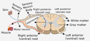
- When a cross-section of the spinal cord is examined, white matter is found on the outside, and gray matter is found inside. The gray matter appears as butterfly-shaped wings within the white matter.
- Two dorsal (posterior) roots emerge from the rear part of the spinal cord. Sensory nerves enter the spinal cord from here.
- Two ventral (front=anterior) roots emerge from the front part of the spinal cord. Motor nerves exit from here.
- The spinal cord also contains interneurons that connect sensory and motor nerves.
Most of the sensory nerves cross over within the spinal cord before reaching the brain.
Basic Functions of the Spinal Cord
- The spinal cord transmits sensory signals from the environment to the brain and conveys the brain’s response to the relevant organs for action.
- It controls habitual movements.
- It manages reflex actions.
- Habits: These are tasks that the brain learns and frequently performs, which are delegated to the spinal cord. Examples include knitting, swimming, riding a bike, dancing, etc.
- If there is any disruption in the execution of these behaviors, the brain intervenes again.
Reflexes are involuntary and automatic movements made in response to a stimulus.
- Reflexes are divided into two types: Innate (Inborn) Reflexes and Acquired (Learned/Conditioned) Reflexes. Reflexes such as the patellar reflex, the infant’s sucking reflex, and the constriction of the pupils in response to light are inherited, innate reflexes.
Acquired reflexes are non-inherited reflexes that are learned through specific training. The center of these reflexes is the brain, and they play a significant role in the development of habits. Behaviors such as swimming, playing the piano, and riding a bike are acquired reflexes learned by the brain and controlled by the spinal cord.
Salivating at the sight of a lemon or a child pulling their hand away from a stove after previously being burned is an acquired reflex. Pulling the hand away from a hot stove is an inherited reflex.
Some innate reflexes are not controlled by the spinal cord. For example, reflexes of the eyes and ears are controlled by the midbrain, while vital internal reflexes such as swallowing, sneezing, coughing, and vomiting are controlled by the medulla oblongata.
The pathway of the reflex, which involves two or more neurons through which the stimulus passes, is called the reflex arc.
The patellar reflex is an inherited reflex. It is the forward movement of the foot when the tendon in the knee is struck. In this simple reflex arc, there are two neurons: one sensory neuron and one motor neuron.
In a complex reflex arc, the sequence involves sensory neurons, interneurons, and motor neurons. The interneuron in this case is located in the spinal cord.
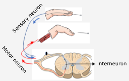
Receptor → Sensory neuron → Dorsal root → Interneuron → Ventral root → Motor neuron → Effector
Reflex movement occurs before the impulse reaches the brain. The sensation of temperature or pain is the result of an impulse being transmitted from the spinal cord to the brain. The impulse is sent through the nerves to the brain, where it is interpreted as a sensation of temperature and pain, leading to voluntary movements. For example, in response to pain, one might think that placing the hand in cold water or blowing on it will reduce the pain.
The spinal cord is controlled by the brain. For example, when we get pricked by a needle, we pull our hand away. However, when a needle is inserted for a blood sample, even if it causes pain, we don’t pull our hand away immediately; we wait. This is because the brain intervenes, interprets, suppresses the reflex, and enables voluntary movement.
A person who gets pricked by a needle will quickly pull their hand away but will feel the pain later. This is because in reflex arcs, impulses are first sent to the spinal cord and then to the brain.
Summary: Functions of Brain Regions
| Brain Region | Subdivision Of | Key Functions |
|---|---|---|
| Cerebrum (Cerebral Cortex) | Forebrain | Voluntary movement, sensory perception, learning, memory, intelligence, consciousness, speech. |
| Thalamus | Forebrain (Diencephalon) | Relay station for sensory impulses (except smell), regulates sleep and wakefulness. |
| Hypothalamus | Forebrain (Diencephalon) | Homeostasis center. Controls body temperature, hunger, thirst, pituitary gland, and autonomic nervous system. |
| Epithalamus | Forebrain (Diencephalon) | Contains the pineal gland (secretes melatonin), regulates biological rhythms. |
| Midbrain | Midbrain | Controls visual and auditory reflexes (e.g., pupil constriction), regulates muscle tone and posture. |
| Cerebellum | Hindbrain | Balance and coordination. Fine-tunes muscle movements, maintains equilibrium. |
| Pons | Hindbrain | Connects cerebellar hemispheres, assists in regulating respiration (breathing control). |
| Medulla Oblongata | Hindbrain | Vital center. Controls heart rate, blood pressure, breathing, and reflexes like swallowing, vomiting, coughing. |